Week 7 - Neuroscience + Art
Week 7 - Neuroscience + Art
 |
| Drawing of Purkinje cells (A) and granule cells (B) from pigeon cerebellum by Santiago Ramón y Cajal, 1899. |
Santiago Ramón y Cajal, the father of modern neuroscience,
is often remembered for his fantastic drawings of neural structures and their
connectivity. Today, his depictions are still used for educational and training
purposes. As mentioned before in week 4’s blog, medicine and art have long been
connected through their concerted efforts to anatomize and document the human
body. Cajal is no exception to this and his illustrations revolutionized the
field of neuroscience, pioneering modern science on neurons and neural
networks.
Art is prevalent in neuroscience even more so when
considering how scientists can visualize microscopic cells. For example, Brainbow
is a process in which individual neurons in a brain sample can be easily
distinguished from neighboring neurons by using fluorescent proteins. Although
the process was developed by professors of Molecular & Cellular biology at
Harvard University, there is no doubt that the brainbows are works of art.
 |
| Brainbow of genetically modified mouse's hippocampus. By Tamily Weissman, Harvard University. |
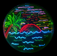
 |
| The Resolution Effect by Amanpreet Badhwar and Pierre Bellec, 2016. |
Today, visual artists and neuroscientists are working together
on developing new, creative techniques for mapping and visualizing the brain. The
Neuro Bureau is a forum and collaborative initiative that hosts conferences,
events and competitions centered around bringing attention to the more aesthetically-orientated
aspects of neuroscience. At one of their exhibitions Reaching Beyond The Obvious,
scientists such as Amanpreet Badhwar and Pierre Bellec develop artwork showing
the aesthetic brilliance of the brain. One of their pieces, The Resolution
Effect, shown at the exhibition in Montreal represents, “the binarized
average functional connectivity matrices generated using functional brain
parcellations of differing sizes (the smaller the parcels,the higher the
resolution). The size of each node is a function of its degree of
connectedness. It alludes to the fact that the dense web of connections
visualized result from the use of high-resolution functional parcellations” (Amanpreet
Cadhwar & Jakobsen).
Sources:
- Badhwar, Amanpreet. Amanpreet
Badhwar. http://www.amanbadhwar.com/.
-
Badhwar, Amanpreet, and
Estrid Jakobsen. “The Interplay between Neuroscience and Art.” Organization
for Human Brain Mapping, 3 June 2017, http://www.ohbmbrainmappingblog.com/1/post/2017/06/the-interplay-between-neuroscience-and-art.html.
-
Frazzetto, Giovanni, and
Suzanne Anker. “Neuroculture.” Nature Reviews. Neuroscience, vol. 10,
no. 11, Nov. 2009, pp. 815–21. PubMed, doi:10.1038/nrn2736.
-
Gangarossa, Giuseppe. “When
Arts Meet Neuroscience...” PLOS Neuroscience Community, 16 Oct. 2016, https://blogs.plos.org/neuro/2016/10/16/when-arts-meet-neuroscience-by-naureen-ghani/.
-
Landau, Elizabeth. “What
the Brain Draws from: Art and Neuroscience.” CNN, 15 Sept. 2012, https://www.cnn.com/2012/09/15/health/art-brain-mind/index.html.
-
“The Neuro Bureau.” The
Neuro Bureau, https://www.neurobureau.org/.
Images:
https://www.ohbmbrainmappingblog.com/blog/the-interplay-between-neuroscience-and-art

Your post made me think about how when we see medical photos they are just that, photos. But when medical professionals look at these photos it truly becomes a piece of dynamic, in depth art piece, source of information, and reassurance.
ReplyDelete